

 en

Hypoxia is a defining feature of many solid tumours and can influence cancer cell proliferation, metabolism, treatment response and metastatic behaviour. However, accurately modelling tumour hypoxia requires careful consideration of how experimental oxygen levels compare with those encountered within tissues and metastatic sites.
In our recent webinar, Diane Lefley, final-year PhD student at the University of Sheffield’s Laboratory for Cancer Metastasis and Dormancy, examined several factors that can influence the interpretation of hypoxia research. Drawing on findings from breast and prostate cancer models, she explored how oxygen concentration, cellular phenotype, hypoxia markers, experimental handling and therapeutic treatment can all affect tumour responses.
At a glance:
- Standard culture conditions do not reflect physiological oxygen levels
- Hypoxia can alter cancer cell proliferation and dormancy
- Hypoxia markers vary between cell lines and conditions
- Chemical hypoxia does not consistently reproduce physical hypoxia
- Reoxygenation can rapidly reduce HIF-1α expression
- Hypoxia can influence therapeutic responses
Why oxygen concentration matters
Standard cell culture conditions can differ considerably from the oxygen environment experienced by cells in the body. Conventional incubators typically use atmospheric oxygen alongside 5% CO₂, resulting in approximately 16% oxygen. This is substantially higher than physiological levels in many tissues. Diane highlighted breast tissue at approximately 5.5% oxygen, prostate tissue at around 4% and bone at approximately 2.9%. Tumour oxygenation can be lower still, depending on factors such as tumour size and proximity to blood vessels. These differences can have significant biological consequences. Hypoxia can affect proliferation, metabolism, DNA damage and repair, while increased reliance on glycolysis can alter lactate production and the surrounding microenvironment. This means that the oxygen concentration used in a cancer model can influence not only cell growth, but also how cells respond to experimental treatments.
Cancer cells do not respond uniformly to hypoxia
Diane's research demonstrated that different cancer cell models can respond very differently to the same oxygen conditions.
At 3% oxygen, MCF7 breast cancer cells continued to proliferate, although more slowly, while MDA-MB-231 cells reached an earlier proliferative plateau. Further comparisons showed that this pattern did not simply correspond to breast cancer receptor status, with other triple-negative adenocarcinoma models continuing to proliferate under hypoxia. The findings suggest that characteristics such as tumour origin and cellular phenotype may be important determinants of the hypoxic response.
Hypoxia also reduced clonogenic potential across the cell lines examined, supporting a possible link between reduced oxygen availability and a more dormant cellular state. This is particularly relevant when modelling metastatic disease, where disseminated tumour cells may remain dormant before progressing to clinically detectable metastases.
Measuring and modelling hypoxia
HIF-1α is widely used to assess cellular responses to hypoxia, alongside markers such as HIF-2α, CA9, GLUT1, VEGF and pimonidazole. However, these markers should not be treated as interchangeable measurements of oxygen concentration. Their expression can also be influenced by cell type, metabolism and other signalling pathways. For example, Diane observed CA9 expression under some nominally normoxic conditions, highlighting the importance of validating hypoxia using appropriate controls and, where possible, complementary markers. The research also examined cobalt chloride as a chemical approach to inducing aspects of the hypoxic response. Its effects varied substantially between cell lines and did not consistently reproduce the responses observed under physical hypoxia. Higher concentrations were also associated with toxicity and changes in cell morphology.
The findings therefore reinforce that chemical activation of hypoxia-associated pathways is not necessarily equivalent to reproducing the physiological effects of reduced oxygen availability.
Reoxygenation can change the experimental picture
An important practical consideration is what happens when hypoxic cells are returned to normal oxygen levels.
Diane found that HIF-1α expression declined rapidly following reoxygenation, although the rate varied between cell lines. In some models, changes could occur within minutes. For experiments involving hypoxic treatment or analysis, this creates a potential source of variation. Removing cells from controlled conditions for procedures such as treatment, imaging or sampling may alter the hypoxia-responsive state researchers are attempting to measure. Maintaining controlled oxygen conditions throughout relevant experimental steps can therefore be important when the aim is to study cells under a defined hypoxic environment.
Hypoxia can influence treatment response
The final part of the research considered how hypoxia may affect responses to cancer treatments, using radium-223 and DNA damage response inhibition as examples. Responses to radium-223 differed between breast cancer cell models under standard and hypoxic conditions, while an ATM inhibitor also showed reduced activity under hypoxia. These differences highlight how oxygen availability can interact with the underlying molecular characteristics of individual tumour models. The research was also extended to an in vivo model of prostate cancer bone metastasis. Here, combining ATM inhibition with radium-223 reduced HIF-1α expression within tumour tissue, while surrounding bone tissue showed a different response. This illustrates the complexity of the tumour microenvironment and the importance of considering how treatments affect both malignant cells and surrounding healthy tissue.
Key Takeaways
- Physiological oxygen concentrations are substantially lower than standard culture conditions.
- Cancer cell responses to hypoxia vary between models and phenotypes.
- Hypoxia markers require validation within the experimental system.
- Chemical hypoxia does not consistently reproduce physical hypoxia.
- Reoxygenation can alter hypoxia-associated signalling within minutes.
- Hypoxia can influence responses to cancer treatments.
Watch the Webinar On-Demand
For a more detailed exploration of the experimental considerations involved in modelling tumour hypoxia, watch the full presentation by Diane Lefley.
Are you looking to put these considerations into practice in your laboratory?
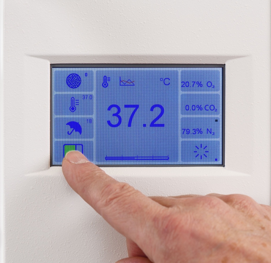
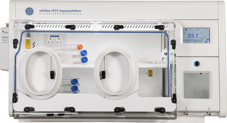
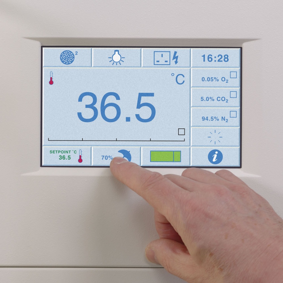
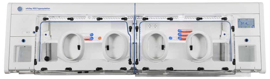
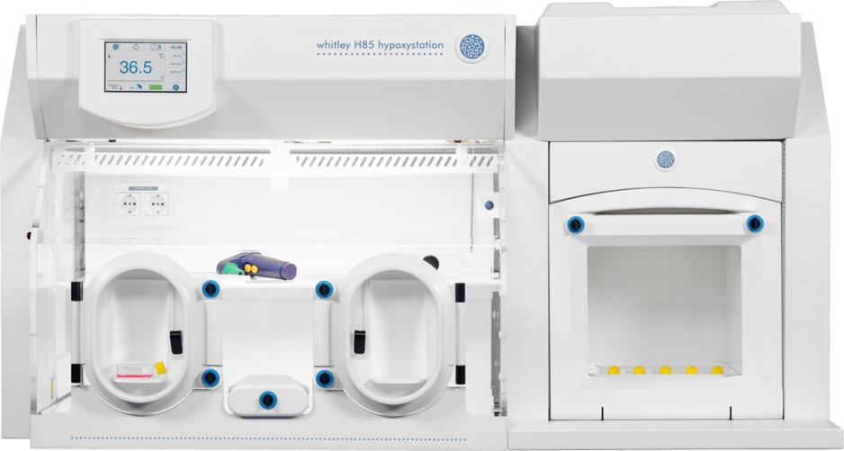
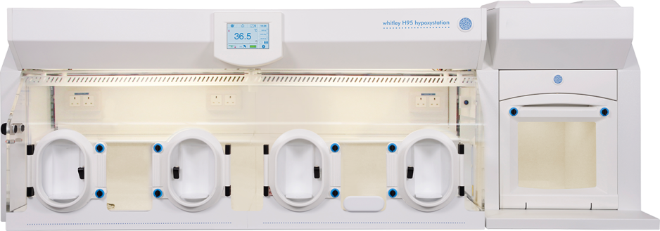
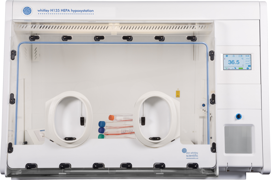
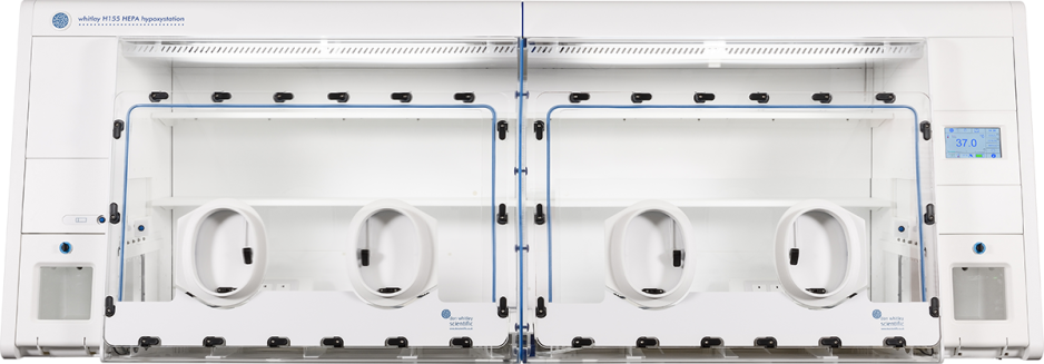
Explore the Whitley Hypoxystation range to accurately and reliably model hypoxic conditions.
The appropriate oxygen concentration depends on the tissue and tumour environment being modelled. While standard cell culture conditions typically result in approximately 16% oxygen, physiological levels are lower, at around 5.5% in breast tissue, 4% in prostate tissue and 2.9% in bone. Tumour oxygen levels can be lower still, so experimental conditions should reflect the biological environment under investigation.
Physical hypoxia involves reducing the oxygen concentration available to cells, whereas chemical hypoxia uses compounds such as cobalt chloride to activate aspects of the hypoxic response. However, chemical hypoxia does not consistently reproduce the effects of reduced oxygen availability and can cause cell toxicity or changes in morphology. Physical hypoxia allows researchers to investigate cellular responses under controlled, reduced-oxygen conditions.
Maintaining controlled oxygen conditions helps researchers avoid changes in hypoxia-associated signalling during experiments. For example, HIF-1α expression can decline within minutes when hypoxic cells are exposed to normal oxygen levels. Reoxygenation during treatment, imaging or sampling can therefore influence results, making it important to consider oxygen exposure throughout relevant experimental procedures.




 England